Total Pageviews

Tuesday, 12 September 2023

CASE 700: RIGHT THORACIC WALL TB ABSCESS, Dr PHAN THANH HẢI, Dr JASMINE THANH XUÂN, Dr HỒ CHÍ TRUNG, Dr LÊ THÔNG LƯU, MEDIC MEDICAL CENTER, HCMC, VIETNAM.

 A 45 year-old  female patient with right thoracic painful swollen area for 5 months.


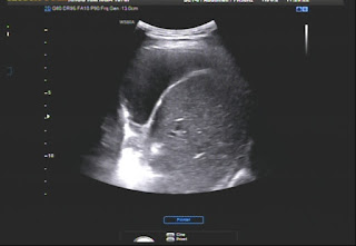
Ultrasound detects right pleural effusion, thoracic wall mass which contains rib cartilage destruction, close by pleural wall thickening at 4 th intercostal space, and local lymph nodes.


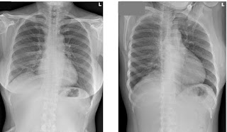
Chest X-RAY  shows right pleural effusion and nothing about thoracic wall. 


MSCT  confirms a right thoracic wall lesion and right lung NAD.


FNAC and core biopsy of right thoracic wall results  think  about TB inflammed lesion with ADA raises slightly in right pleural fluid.


So it exists a painful right thoracic wall for 5 months and evidents belongs to a TB infection without primary lung lesion.


Histoimmumologic staining results are TB inflammed cartilage and soft tissue which exists granular cells and lymphocytes.

It will be planned for a TB regimen in TB and Lung hospital.

Thursday, 7 September 2023

CASE 699: DIFFUSE LARGE B CELL LYMPHOMA and WALDEYER'S RING, Dr PHAN THANH HẢI, Dr DƯƠNG XUÂN TÙNG , MEDIC MEDICAL CENTER, HCMC, VIETNAM

A male patient 41 year-old with multiple cervical nodes and sore throat as tonsillitis.


Chest X-RAY notes left lung lesion.


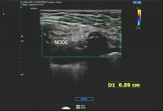
Ultrasound detects many lymph nodes  # 17-15-13 mm without nodal hilus, solid, hypoechoic on his neck and in the abdomen : at liver hilus :27mm, mesenteric,  pelvic: 17-21 mm and a splenomegaly :141mm. Results of ultrasound notes a multiple lymph nodes in cervical, supraclavicular and abdominal region that leads to a diffuse lymphoma. 


Biopsies of tonsils and pharyngeal cavum results are lymphoma infiltration without immunohistochemical staining.


ENT examinations results are many lesions of tonsils and oral cavity and Waldayer's ring.


Biopsy of right tonsil ulcer for ruling out cancer and immuohistochemical staining result is diffuse lymphoma type large B cell.


Patient goes through a chemotherapy planning for lymphoma. Cervical nodes reduce their sizes with effective management. 


REFERENCES:


Friday, 1 September 2023

CASE 698: HTA YOUNG PATIENT with 2 RENAL ARTERIES each side, Dr PHAN THANH HẢI, Dr PHAN THANH HẢI PHƯỢNG, Dr HỒ KHÁNH ĐỨC, MEDIC MEDICAL CENTER, HCMC, VIETNAM

Ultrasound feels uncertainly about a  50% stenosis of renal artery which its diameter is # 7.5 milimeter.
Renal structures and sizes seem being in normal limits.


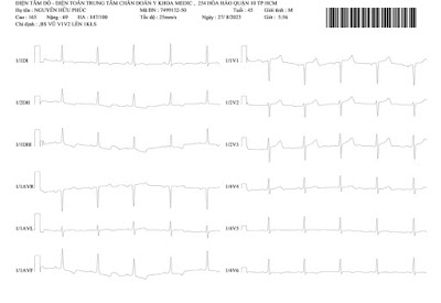
Chest X-RAY film is normal and his EKG results ischemic heart disease.


Lab data shows renal insufficiency: eGFR 37mL/min/1.73m2; creatinin 2.2mg/dL;  and hyperuricemia with serum urea 65.84mg/dL


At last, DSA detects 2 renal arteries each side that belongs to a renal artery malformation (bilateral duplication of renal artery).


So the male patient with renal duplicated arteries might be suffering from parenchymal nephropathy /and due to gout.


REFERENCE


Renal arteries are a pair of lateral branches from abdominal aorta. Normally each kidney receives one renal artery. However, accessory renal arteries can also exist. The normal renal arteries enter the kidney through its hilum where as the accessory renal arteries might enter the renal artery through the hilum or through the surfaces of the kidney. Knowledge of the variations in the renal arteries is important for urologists, radiologists and surgeons in general.


Accessory renal arteries are common in 20–30% of individuals, usually arising from the aorta above or below the main renal artery. The variation in the number of arteries is because of persistence of lateral splanchnic arteries or due to the persistence of blood supply from lower level than normal.


Tuesday, 29 August 2023

CASE 697: STOMACH GIST, Dr PHAN THANH HẢI, Dr PHAN THỊ HƯỜNG, MEDIC MEDICAL CENTER, HCMC, VIETNAM.
 A 47 year-old female patient with a gastric tumor at lesser curvature which was incidentally revealed by ultrasound.

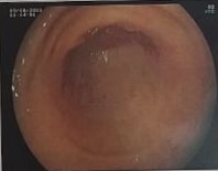
But gastroendoscopy result was only a gastritis.


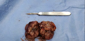
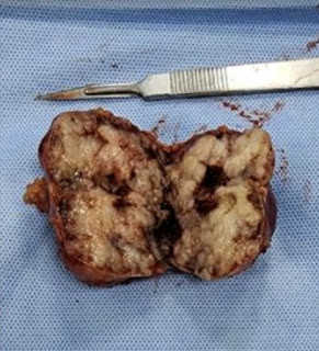
Later MSCT confirmed a 50x58 mm gastric wall tumor maybe a stomach GIST. The tumor captured CE inhomogeneously and grew out of its way to compress the other organs around in abdominal cavity.


Robot surgery removed entirely the gastric tumor and recreated the cardia. The histopathological result is gastric GIST.


REFERENCE:


A 53 yearale patient with a gastric tumor which is spontaneously evealed 


Tuesday, 22 August 2023

CASE 696: PRIMARY CLEAR CELL HCC, Dr PHAN THANH HẢI, Dr NGÔ THỊ HUYỀN TRANG, MEDIC MEDICAL CENTER, HCMC, VIETNAM


A male patient 53 year-old with tumor in right lobe of liver and negative WAKO tests.

A 55 x 42 mm liver tumor is observed on ultrasound in segment 7. It is almost uniform, well-limited, weakly vascularized, and exhibits elastography ultrasonography SWE that is five times harder than hepatic parenchyma, measuring 29 kPa as opposed to 6.3 kPa. HCC risk testing is negative with WAKO.


MRI with Primovist confirms a 50 milimeter clear cell HCC (CC-HCC). T2 CE captured signals are higher than liver parenchyma and  lower than on T1. 


Biopsy results of tumor is an HCC well differentiazed.


REFERENCE:


Hepatocellular carcinoma (HCC) is a common cancer world-wide with a higher incidence in Asia. Clear cell variant of HCC (CC-HCC) has a frequency ranging from 0.4% to 37%. The presence of 90-100% clear cells is rare.