Total Pageviews

Thursday, 3 August 2023

CASE 693: ADVANCED MALIGNANT MESENCHYMAL CELLS of PROSTATIC TUMOR, Dr PHAN THANH HAI, Dr NGUYEN MINH THIEN, MEDIC MEDICAL CENTER. HCMC, VIETNAM.

 A 66 year-old male patient with weight loss and asthenia. Some 15-30 mm metastasized necrotic periaortic lymph nodes and prostate hypertrophy are noted on abdominal ultrasound.  Chest X-ray is normal and abdominal MSCT confirmes retroperitoneal lymph nodes and prostate tumor. PSA value is 1.38 ng/mL but with microscopic blood urine that transfers him to Medic Center for a prostate biopsy.

By via TRUS it exists a #55x43x62 mm big prostate, loss its capsule and distorsion of prostatic structure. The prostate tumor is noted having of many hard sites on shear wave ultrasound elastography.


Performed 12 specimen prostate biopsy and 16 items histoimmunopathologic report concludes a non- specialized mesenchymal cell of prostate tumor (PCa) on a chronic TB inflamed based structure.


This is a rare PCa malignant mesenchymal cell  of prostate tumor may happen on 1/1000 cases. The patient is waiting for an appropriate management due to his advanced status of the tumoral progress.


Thursday, 27 July 2023

CASE 692: Five Year HCC post-op, Dr PHAN THANH HẢI, Dr DƯƠNG NGỌC THÀNH, MEDIC MEDICAL CENTER, HCMC , VIETNAM.

 A 71 year-old female patient with her history of a liver tumor which was quite difficult diagnosed 5 years before.

She had been go through ultrasound, MSCT  and liver biopsy 2 times in 2 other hospitals, but the results were benign hepatic tumor and no clue of infected HCV and HBV.

In Medic Center, ultrasound noted a 50 milimeter right liver hemangioma and MRI  with gado was an AML but not ruling out HCC.


But HCC risk tests with WAKO test were positive and positive antiHBe in that time.


After 2 months of the five year before to examine the liver tumor, the HCC  diagnosis was made and the tumor was removed surgically when the patient aged 66.


Histopathological report was a poor differentiazed carcinoma of liver with fatty degeneration  and hepatic chronic inflammation.

And 5 years went by the female patient remains well. The right lobe of liver exists a fibrosis scar.


The prognosis of HCC patient will be well if an effective management performs successfully in time for the female patient [Acceptable 5-year survival].

Saturday, 15 July 2023

CASE 691: LEFT ADRENAL TUMOR, Dr PHAN THANH HẢI, Dr VÕ THỊ PHƯƠNG TRINH, MEDIC MEDICAL CENTER, HCMC, VIETNAM


A 48 year-old female patient without arterial hypertension signs is incidentally detected a tumor of adrenal gland #20×22 mm by ultrasound.


MSCT confirmes  a 16x22 mm left adrenal benign tumor  which HU before CE is 69 and HU post CE =26. CE discharge is over 50% that may proove the benignity of the tumor.


4 months after the left adrenal tumor is removed by endoscopic surgery.


Result of pathology is a benign adrenal tumor with thin fibrous capsule.


Thursday, 13 July 2023

CASE 690: APPENDICULAR ABSCESS, Dr PHAN THANH HAI, Dr NGUYEN NGOC XUÂN GIANG, MEDIC MEDICAL CENTER - BINH AN HOSPITAL KIÊN GIANG, VIETNAM

A 56 year-old female patient with umbilical pain for one week, comes to Binh an hospital after 4 days in constipation.

Ultrasound detects an abscess in RLQ but could not rule out a PID. Although sonologist notes an appendicular abscess but could not reveal the inflammed appendix into the pelvic abscess.


MSCT confirms a 60x80 mm  right pelvic abscess contrast captured and edema of the wall of terminal jejunum. The report is pelvic abscess maybe an appendicular abscess.


Endoscopic surgery drained the pelvic abscess and the patient remains well.


Thursday, 6 July 2023

CASE 689: RECTUM WALL GIST, Dr PHAN THANH HẢI, Dr LÊ TUẤN KHUÊ, MEDIC MEDICAL CENTER, HCMC, VIETNAM

A 56 year-old female patient with 6 month constipation and bleeding from her anus  for 2 weeks.
Rectal endoscopic result is a hemorrhagic rectal tumor which is far from anal orifice 20 millimeters.

MRI confirms a 53x41 mm tumor of the rectal canal.


A transperineal biopsy with ultrasound-based guiding is done for a 42x48x44mm vascularised solid mixed tumor.


Result of histoimmunology is a GIST tumor of  rectum wall.


The female patient is planned for a surgery and chemotherapy.

Saturday, 17 June 2023

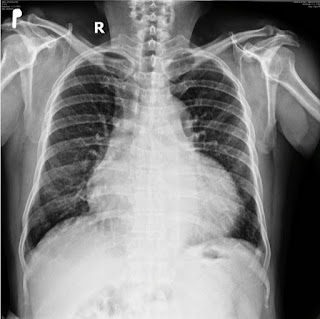
CASE 688: ASYMMETRICAL HYPERTROPHY of the HEART, Dr PHAN THANH HẢI, Dr NGUYỄN DINH, Dr ĐÀO XUÂN DUNG,MEDIC MEDICAL CENTER, HCMC VIETNAM.


A 59 year-old arterial hypertension male patient with a chest pain in taking alcohol from October 2019. He came Heart Institut HCMC with a note of infarctus of myocardium from Cantho province.  


But MSCT in Hoan my hospital Cantho province proves a slight stenosis 30% of coronary arteries. 

In COVID 19 pandemic, he goes around the private clinics with his HTA status and his heart but nothing change.

In May to July 2022 he goes to Medic Can tho and then Medic Hoa Hao Center in HCMC.

An coronary CTA of this second time shows slight stenosis under 30% and slight pericardial effusion but notes a hypertrophic cardiomyopathy of his heart.


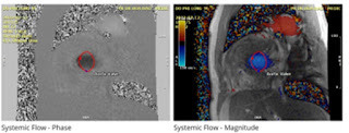
At last Cardiac MRI confirms an asymmetrical hypertrophy of the heart.


He gets well with the management of  hypertrophic cardiomyopathy.


REFERENCE


Thursday, 15 June 2023

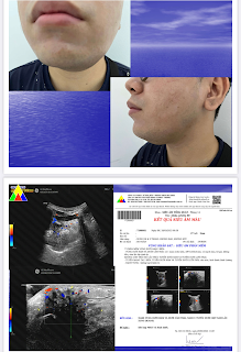
CASE 687: SUBMANDIBULAR TUMOR, Dr PHAN THANH HẢI, Dr TRẦN LÃM, MEDIC MEDICAL CENTER, HCMC, VIETNAM


 A 27 year-old male patient suffers from left submandibular gland for 3 years.

Ultrasound notes a left submadibular gland tumor.


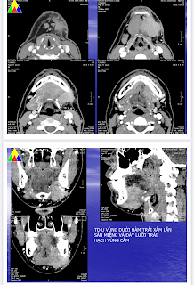
MSCT confirms the left submandibular  that invaded the mouth floor and node.


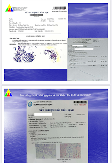
But result of FNAC has only an amount epithelium cells and RBC.  And in the second time maybe a pleomorphic tumor.

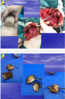
Goes through a surgery, the specimen is a malignant submandibular gland tumor, mucoepidermoid carcinoma low-grade.