Total Pageviews

Thursday, 13 April 2023

CASE 678: COMPLICATED DIVERTICULITIS, Dr PHAN THANH HẢI, Dr LÊ THỊ THANH THẢO, MEDIC MEDICAL CENTER, HCMC, VIETNAM.

A 55 year-old female diabetic patient suffers from epigatric pain crisii for 4 days and diarrhea.

Ultrasound detects an amount of abdominal free fluid,  two RLQ abscesses, and edema of peritoneum.


MSCT confirms the peritonitis due to 2 abscesses at the right flank of the abdomen.


A right colectomy is performed as perforation of colonic diverticulitis # 2x2 cm of transverse colon and the other one of cecum.

Subhepatic drainage is made to withdraw fluid out and an artificial anus is done.

 And the female patient remains well.


Wednesday, 12 April 2023

CASE 677: LUNG TUMOR, Dr PHAN THANH HẢI, Dr VÕ NGUYỄN THỤC QUYÊN, MEDIC MEDICAL CENTER, HCMC, VIETNAM.

A 53 year-old female patient with left thoracic pain for some months. She went through 2  lung tumor (hemangioma) surgeries respectively for 15 years  in K hospital and 4 years (Binh Dan hospital) before.


Pulmonary function report is normal.


Ultrasound notes a round mass  #50x39 mm at the bottom of left lung. It is a cystic lesion, well-limited capsule, and  belongs to the soft code on elastography ultrasound.


MSCT confirms a left pulmonary septated cyst  # 45x29 mm with sludge.


Surgery in the third time (on Tuesday 12, April, 2023) performs for removing the left lung cystic lesion. 

Gross specimen of the left lung tumor.


Microscopic result is malignant hemangiopericytoma.


Monday, 10 April 2023

CASE 676: LUNG TUMOR or TB LUNG, Dr PHAN THANH HẢI, Dr LÊ HỮU LINH, MEDIC MEDICAL CENTER, HCMC, VIETNAM.


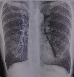
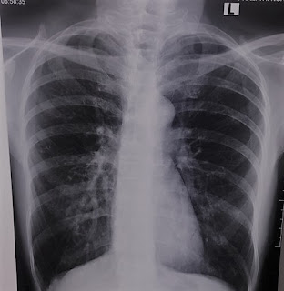
 A 57 year-old female patient with a left lung (lingula) mass on chest X-Ray film on January 2023 without any symptom.


But its size changes for months on the chest X-ray film (Mars 2023).


Lung ultrasound notes a consolidation region #44x31mm of left lower lobe of lung, with air bronchogramme, and soft code of elastography ultrasound (ARFI technique).


Chest MSCT later confirms the 50 mm left lung mass and biopsy.

Result of biopsy is a TB lung mass with TB cyst, caseous necrosis,  lymph cells  and Langhans cells.


The female patient then starts a TB regimen. 


Saturday, 25 March 2023

CASE 675: SCHWANNOMA of C5 ROOT of LEFT BRACHIAL PLEXUS, Dr PHAN THANH HẢI, Dr LÊ THỊ THANH THẢO, MEDIC MEDICAL CENTER, HCMC, VIETNAM.

A  56 year-old female patient  with left facial numbness [?] for 3 days.

Neck ultrasound at left supraclavicular region detects # 8-19 mm hypoechoic enlargement of left brachial plexus maybe due to schwannoma or neurinoma. While ultrasound examines at the neck region the female patient feels her left upper arm numb sensation (due to affecting the left upper brachial nerves from C 5 root).


MRI confirms a schwannoma tumor #21x12mm of left C 5 root.


Surgery removes the tumor of C 5 root from left brachial plexus which the pathohistology result is the schwannoma of the C5 root.


REFERENCE


Thursday, 23 March 2023

CASE 674: BREAST SARCOMA, Dr PHAN THANH HẢI, Dr LÊ THÔNG LƯU, Dr JASMINE THANH XUÂN, MEDIC MEDICAL CENTER, HCMC, VIETNAM.

 A  61 year-old female patient  with right breast tumor and axillary nodes.

Breast ultrasound detects a  # 28x10 mm mass of the right breast BI-RADS 4 B and second tumor of this one, #19×10mm. And elastography ultrasound notes a hard code of it.


Breast MRI represents 2 right breast tumors, BI-RADS 5.


Biopsy of right axillary node is inflammed node, but result of core biopsy of the right breast tumor is poor differentiated breast sarcoma (C 49).


Total right mastectomy is performed,  and the final results are the right breast sarcoma and the chronic inflammed axillary node.

Saturday, 18 March 2023

CASE 673: RENAL INFARCTION, Dr PHAN THANH HẢI, Dr NGUYỄN NGHIỆP VĂN, MEDIC MEDICAL CENTER, HCMC, VIETNAM.

A 50 year-old HTA female patient  with left flank pain, headache and red blood cells in urine for 3 days.


Ultrasound detects a 93x43 milimeter hypoechoic left kidney  without Doppler signal via SMI technique while the right one  110x47 mm is normal.


Elastography of left kidney SWE is in hard code.


MSCT Angio represents left kidney atresia as thrombosis of the left renal artery which leads it to be total obstruction.


It’s a left renal infarction case which is in late inspection.

Thursday, 16 March 2023

CASE 672: HYDATID CYST in MYOCARDIUM, Dr PHAN THANH HẢI, Dr TRẦN THỊ MINH PHƯỢNG, MEDICAL MEDICAL CENTER, VIETNAM.

A  75 year-old female patient  with asthenia and dyspnea for one year. The eosinophil  proportion of white blood cell is of 0.14 and Echinococcus (hydated) IgM Elisa negative is of 0.16 index.

Cardiac ultrasound detects a 30 mm simple cyst (Gharbi type I  hydatid cyst) in myocardium at the cardiac apex.


MSCT and MRI confirm the cardiac cyst in myocardium at the apex of the heart.


Surgery removes successfully the hydatid cyst with larva from heart. 


Microbioparasitology result is hydatid cyst with larva of Echinococcus sp.


Hydated cyst in myocardium a very rare site coming over that usually appears in liver, spleen, lung ... instead.

Sequencing result is Echinococcus ortleppi.