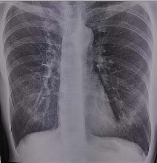
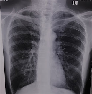
A 57 year-old female patient with a left lung (lingula) mass on chest X-Ray film on January 2023 without any symptom.
But its size changes for months on the chest X-ray film (Mars 2023).
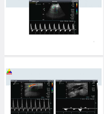
Lung ultrasound notes a consolidation region #44x31mm of left lower lobe of lung, with air bronchogramme, and soft code of elastography ultrasound (ARFI technique).
Chest MSCT later confirms the 50 mm left lung mass and biopsy.
Result of biopsy is a TB lung mass with TB cyst, caseous necrosis, lymph cells and Langhans cells.
The female patient then starts a TB regimen.


.jpg)
.jpg)
.jpg)

.jpg)
.jpg)
.jpg)
.jpg)
.jpg)
.jpg)
.jpg)
.jpg)
.jpg)
.jpg)
.jpg)
.jpg)
.jpg)
.jpg)
.jpg)
.jpg)
.png)
.jpg)


