Total Pageviews

Thursday, 23 March 2023

CASE 674: BREAST SARCOMA, Dr PHAN THANH HẢI, Dr LÊ THÔNG LƯU, Dr JASMINE THANH XUÂN, MEDIC MEDICAL CENTER, HCMC, VIETNAM.

 A  61 year-old female patient  with right breast tumor and axillary nodes.

Breast ultrasound detects a  # 28x10 mm mass of the right breast BI-RADS 4 B and second tumor of this one, #19×10mm. And elastography ultrasound notes a hard code of it.


Breast MRI represents 2 right breast tumors, BI-RADS 5.


Biopsy of right axillary node is inflammed node, but result of core biopsy of the right breast tumor is poor differentiated breast sarcoma (C 49).


Total right mastectomy is performed,  and the final results are the right breast sarcoma and the chronic inflammed axillary node.

Saturday, 18 March 2023

CASE 673: RENAL INFARCTION, Dr PHAN THANH HẢI, Dr NGUYỄN NGHIỆP VĂN, MEDIC MEDICAL CENTER, HCMC, VIETNAM.

A 50 year-old HTA female patient  with left flank pain, headache and red blood cells in urine for 3 days.


Ultrasound detects a 93x43 milimeter hypoechoic left kidney  without Doppler signal via SMI technique while the right one  110x47 mm is normal.


Elastography of left kidney SWE is in hard code.


MSCT Angio represents left kidney atresia as thrombosis of the left renal artery which leads it to be total obstruction.


It’s a left renal infarction case which is in late inspection.

Thursday, 16 March 2023

CASE 672: HYDATID CYST in MYOCARDIUM, Dr PHAN THANH HẢI, Dr TRẦN THỊ MINH PHƯỢNG, MEDICAL MEDICAL CENTER, VIETNAM.

A  75 year-old female patient  with asthenia and dyspnea for one year. The eosinophil  proportion of white blood cell is of 0.14 and Echinococcus (hydated) IgM Elisa negative is of 0.16 index.

Cardiac ultrasound detects a 30 mm simple cyst (Gharbi type I  hydatid cyst) in myocardium at the cardiac apex.


MSCT and MRI confirm the cardiac cyst in myocardium at the apex of the heart.


Surgery removes successfully the hydatid cyst with larva from heart. 


Microbioparasitology result is hydatid cyst with larva of Echinococcus sp.


Hydated cyst in myocardium a very rare site coming over that usually appears in liver, spleen, lung ... instead.

Sequencing result is Echinococcus ortleppi.

Saturday, 11 March 2023

CASE 671: BetaHCG SECRETION by a PULMONARY CARCINOMA, Dr PHAN THANH HẢI, Dr NEANG SI PHATS, Dr HUỲNH TRÁC LUÂN, MEDIC MEDICAL CENTER, HCMC, VIETNAM.

A  42 year-old female patient,  TPAL 1001 with critical menorrhegia for one month  and weight loss and anemia. HTA:100/70 mmHg, PA:110 bpm. W = 40 kg H: 155mm, BMI: 16.6.

TVS ultrasound detects only uterine fibroma, but beta HCG highly rising= 69,474 mlU/mL.


Total body MRI detects a 98 mm right lung tumor and a 25 mm uterine fibroma.


Chest X-Ray film represents tumor of the right lung which is thought a beta HCG secretion tumor.


Lung biopsy result is a non small cell adenocarcinoma, stage IIIc, invasive at lung.


As the poor status of the patient, the current clinical management is blood transfusion to improve her life in waiting for a reexamination in a cancer hospital.

REFERENCE:


Saturday, 25 February 2023

CASE 670: CHRONIC AORTIC DISSECTION, Dr PHAN THANH HẢI, Dr TRẦN THANH NGA, Dr VÕ NG THÀNH NHÂN, MEDIC MEDICAL CENTER, HCMC, VIETNAM

A 62 year-old HTA female patient with asthenia, impermanent chest pain for one month, is diagnosed aortic dissection by color Doppler ultrasound. She has got slightly leg edema for one week before ultrasound examination.

HTA: 150/80 mmHg, HR : 115 b/min.

Chest X-ray shows pneumonia of upper lobe of the right lung without pleural effusion.


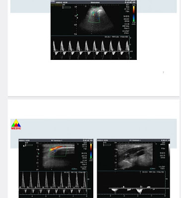
Ultrasound detects a thick flap inside the abdominal aortic lumen which separates the 22 milimeter aorta into 2 colored Doppler code lumens. The aortic dissection represents from thorax to right iliac artery. Bloodstreams in two lumens of the aorta are different with one velocity of 66 cm/s lower than the other, 166 cm/s. 


AngioMSCT confirmes aortic dissection from aortic arc in the chest to iliac artery in thr abdomen. The diameters of aorta are ascending 35mm, aortic arc, 33mm, abdominal, 30 mm respectively. The left kidney artery comes from the lower velocity lumen.


Stenting the aortic dissection is the appropriate management for the chronic aortic dissection w
and antihypertensive treatment.

REFERENCE


Thursday, 23 February 2023

CASE 669: PROSTATIC CYST or A-V FISTULA, Dr PHAN THANH HẢI, Dr LÝ THANH LÃM, Dr LÊ THANH LIÊM, Dr VÕ NG THÀNH NHÂN, Dr HỒ KHÁNH ÐỨC, MEDIC MEDICAL CENTER, HCMC, VIETNAM

A 62 year-old male HTA patient with an aliasing cystic lesion close by his prostate without urogenic symptom was revealed by Doppler technique. A note of an pseudoaneurysm of the right internal iliac artery due to A-V fistula is written down, but it has been missed out on it in 3 other examinations in the past.


MSCT and Angio confirm the pseudoaneurysm of right internal iliac artery.


After a vascular collation via pelvic angiography the pseudoaneurysm disappeares.


Without Doppler technique we could be in error or misdiagnose some cystic lesion.

Saturday, 18 February 2023

CASE 668: THYROID TUMOR, Dr PHAN THANH HẢI, Dr PHẠM THỊ THANH XUÂN, MEDIC MEDICAL CENTER, VIETNAM.

A 64 year-old male diabetic patient with right painfull swollen neck for 2 weeks, he has also got impermanent tinnitus and hoarse voice due to right vocal cord paralysis.


Ultrasound of his neck shows a 29x33 mm big right lobe of thyroid, hypoechoic mixed pattern, poor-limited while the left lobe is intact with its slightly small size #15x17mm. It exists many round and ovale, hypoechoic lymph nodes on both 2 sides of his neck maybe due to metastase or infiltrated lymphoma.


Result of lymph node biopsy is poorly differentiated carcinoma metastase node.


MSCT of neck and chest detect lymph nodes in mediastinum, and on 2 sides of neck that are thought due to metastasis from unknown origin. The  right lobe of thyroid represents undetermined lesion and low captured CE. 


As a poorly differentiated carcinoma of thyroid is a rare entity, a FNAC of right thyroid lobe is performed.


And the result of FNAC of right lobe of thyroid is poorly differentiated thyroid carcinoma.

As the poor status the patient just only undergoes a chemotherapy.