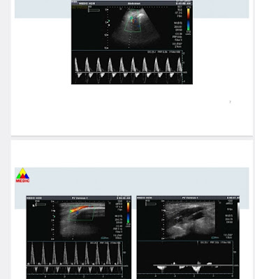
A 62 year-old HTA female patient with asthenia, impermanent chest pain for one month, is diagnosed aortic dissection by color Doppler ultrasound. She has got slightly leg edema for one week before ultrasound examination.
HTA: 150/80 mmHg, HR : 115 b/min.
Chest X-ray shows pneumonia of upper lobe of the right lung without pleural effusion.
Ultrasound detects a thick flap inside the abdominal aortic lumen which separates the 22 milimeter aorta into 2 colored Doppler code lumens. The aortic dissection represents from thorax to right iliac artery. Bloodstreams in two lumens of the aorta are different with one velocity of 66 cm/s lower than the other, 166 cm/s.
AngioMSCT confirmes aortic dissection from aortic arc in the chest to iliac artery in thr abdomen. The diameters of aorta are ascending 35mm, aortic arc, 33mm, abdominal, 30 mm respectively. The left kidney artery comes from the lower velocity lumen.
Stenting the aortic dissection is the appropriate management for the chronic aortic dissection w
and antihypertensive treatment.
REFERENCE
.jpg)
.jpg)
.jpg)
.jpg)
.jpg)
.jpg)
.jpg)
.jpg)
.jpg)
.png)
.jpg)


.jpg)


.jpg)
.jpg)
.jpg)
.jpg)
.jpg)
.jpg)
.jpg)
.jpg)


.jpg)
.jpg)
.jpg)
.jpg)
.jpg)
.jpg)
.jpg)