Total Pageviews

Thursday, 9 February 2023

CASE 667: LEFT KIDNEY UTUC, Dr PHAN THANH HẢI, Dr TRẦN THANH NGA, MEDIC MEDICAL CENTER, VIETNAM


A 51 year-old male patient with hematuria for 6 months but since last month he suffered from a full hematuria. Ultrasound detected a hypoechoic mass nearby left inferior renal pole. SMI showed hypervascular pattern inside the mass.

MSCT later confirmed a left renal tumor RCC or oncocytoma and a proximal upper left ureter tumor.

Surgery removed the left kidney, left ureter and a part of bladder as high malignant upper tract urothelial cancer (UTUC).


Saturday, 4 February 2023

CASE 666: GIANT HYDRONEPHROSIS, Dr PHAN THANH HẢI, Dr TRẦN NGÂN CHÂU, MEDIC MEDICAL CENTER, HCMC, VIETNAM

 A 53 year-old male patient with a painless big abdomen was accidentally detected having a right giant hydronephrosis via ultrasound examination. 


As the right dilated kidney was in large size that could not use any classic section to find out the clue of the renal obstruction. But in going down  from the left pelvis and toward right side, ultrasound revealed an evident ureteral stone # 20 milimeter.


Later, MSCT confirmed easier than ultrasound the 20 mm right ureteral stone which caused the right giant hydronephrosis.


The patient went through a right nephrostomy in emergency situation. About 4.5 liters of urine was drained out and then his abdomen getting flatten.

Two weeks later was done an evaluation of the right kidney function via Tc-99m DTPA scan. Ultrasound re-examination noted a distortion of right kidney structure: thickness of renal cortex thinner than 6 millimeter and nonexistent differentiation of renal medulla from cortex of renal parenchyma.


Via endoscopy  right nephrectomy was performed as the right disfunction kidney. 


6 months later value of eGFR rised from 64 to 75 mL/min/1.73m2 , and the patient remains well.

Tuesday, 17 January 2023

CASE 665: INTESTINAL GIST, Dr PHAN THANH HẢI, Dr TRƯƠNG THỊ NGỌC TIẾN, MEDIC MEDICAL CENTER, HCMC, VIETNAM

 A 55 year-old female patient suffers from a painful epigastric mass for one week. As gastritis management failed, ultrasound reexamines her abdomen.

A 117x67 milimeter mass is detected between liver and right kidney. Its structure is poor with some cystic lesions and central part more rich echogeneicity that is thought to be an intestinal GIST. 

Her uterus gets having some fibroma lesions also which have the same echogeneicity.


MSCT later confirmes the intestinal GIST and uterine fibroma.


Surgery removes the intestinal GIST [left specimen] and an uterine fibroma with stalk (right specimen).


The histopathologic and chemohistoimmuno- staining results are intestinal GIST, and uterine leiomyoma.


Thursday, 5 January 2023

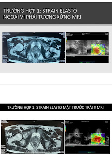
CASE 664: 03 CASES of PROSTATE DISORDERS with LOW VALUES of PSA, Dr PHAN THANH HAI, Dr NGUYEN MINH THIEN, MEDIC MEDICAL CENTER, HCMC, VIETNAM.

 3 cases of prostate disorders wit low values of PSA= two cases of TB and one case of carcinoma that concludes prostate cancer may happen with any value of PSA [but not only PSA>4 ng/mL]; although value of PSA being considered a guiding lab data for prostate cancer.

Should base on free PSA, DRE, MRI, elasto ultrasound together for screening prostate cancer, but these lab data and diagnostic imaging not stand for a diagnostic of prostate cancer.

CASE 1


CASE 2


CASE 3:

Chief complaint: only left swollen scrotum.


Friday, 23 December 2022

CASE 663: ENDOMETRIOSIS and DIENOGEST, Dr PHAN THANH HAI, Dr NGUYEN THI XUAN HOA, MEDIC MEDICAL CENTER, HCMC, VIETNAM

A 38 year-old female patient, TPAL 1-0-0-1, with sideropenia, dysmenorrhea and menorrhagia that has been diagnosed endometriosis in uterus and right ovary by transvaginal ultrasound  (TVUS) and pelvic MRI.

On MRI, size of endometriosis in uterus # 5×7 centimeter and, in ovary # 1.5 centimeter.


She is managed for iron deficiency, and her endometriosis with Dienogest 2mg one tablet per day. For 5 months, focal lesions in her uterus and right ovary are getting smaller sizes. Dysmenorrhea appears slightly and she is with the new generation of progestin acceptable menorrhagia.


NOTA:


Thursday, 22 December 2022

CASE 662: MYOCARDIAL INFARCTUS with ST non Elevated, Dr PHAN THANH HAI, Dr BUI BINH HUAN, Dr VU BICH DOAN TRANG , MEDIC MEDICAL CENTER, HCMC, VIETNAM

 A 68 year-old HTA male patient with right chest pain for 8 hours while holding some water buckets.

Troponin I: 648.6 ng/L, CPK MP: 13.32 ng/mL are elevated, but EKG and cardiosonography have no clue of an acute myocardial infarctus.


About 50% patients of unstable angina  [UA] and NSTE MI have no change of EKG in early time while troponin-I elevated during the first 24 hours.

Based on elevated troponin-I and CPK, the patient is transferred to an emergency hospital after a diagnosis of myocardial infarctus is made.

Saturday, 17 December 2022

CASE 661: RCC DETECTED in Check-up ULTRASOUND, Dr PHAN THANH HAI, Dr NGUYEN PHUOC TOAN, MEDIC MEDICAL CENTER, HCMC, VIETNAM

A 45 year-old male employee is detected occasionally a # 25 mm left renal tumor in a check-up by ultrasound in his company. He has not signs of lumbago and hematuria.


MSCT later confirms the 30 mm left kidney tumor at lower pole and anterior face which is thought maybe a RCC.


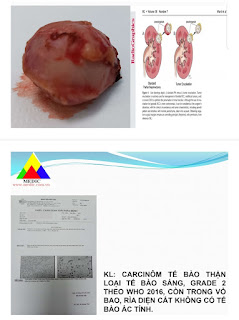
Endoscopic surgery removes the tumor in its capsule by pure enucleation technique.


Result of histopathogy is a clear cell renal carcinoma, grade 2.

Portable ultrasound in control check-up for worker and employee in the companies may detect some cases with subclinical symptom. If it exists any abnormal evident, they will be called going to our center for more informations by other modalities of diagnostic imaging and added lab data. With a final clear diagnosis he/she will be help by an available management.