Total Pageviews
Thursday, 20 October 2022
CASE 654: PHYLLODES TUMOR of the BREAST, Dr PHAN THANH HẢI, Dr JASMINE THANH XUÂN, Dr TRẦN THỊ HỒNG VÂN, MEDIC MEDICAL CENTER, HCMC, VIETNAM.

Thursday, 13 October 2022
CASE 653: PRIMARY MUSCLE LYMPHOMA, Dr PHẠM THỊ THANH XUÂN, Dr PHAN THANH HẢI, MEDIC MEDICAL CENTER, HCMC, VIETNAM
A 68 year-old male patient with a mass at right neck in lung TB regimen for 4 months but still weight loss and sudation. The painful mass existed for 1 month and getting bigger with skin redness.
Soft tissue ultrasound detected a complexe mass #57x43 mm in muscle at right neck from angula of lower maxillary region which distorsed structure, with intramuscular fluid beside cervical vertebral column C4. It existed not any neck lymph node.
MRI confirmed a right neck tumor invasive to muscle.
Chest CT = no lung invasive, no mediastinal lymph node nor axillary node. Bone marrow biopsy exist not any malignant cell.
In surgical biopsy for chemohistopathology of the tumor resulted small cell lymphoma (C83).
The patient was treated TB lung completely and then continued lymphoma chemotherapy. Now the muscular tumor was smaller 80% and the patient remains well.
Primary muscle lymphoma is very rare entity without characteristic imaging findings but diagnostic imaging keeps a role.
REFERENCES:
Cancer Imaging (2013) 13(4), 448457 DOI: 10.1102/1470-7330.2013.0036
Imaging of musculoskeletal lymphoma
https://www.leukaemia.org.au/blood-cancer-information/types-of-bloodcancer/lymphoma/non- hodgkin-lymphoma/small-lymphocytic-lymphoma/
https://www.cancersupportcommunity.org/chronic-lymphocytic leukemiasmalllymphocytic-lymphoma
https://patientpower.info/the-curious-case-of-cll-and-sll-leukemia-lymphoma-orboth/
https://www.ncbi.nlm.nih.gov/pmc/articles/PMC6400341/
https://ashpublications.org/blood/article/131/25/2745/37141/iwCLL-guidelines-fordiagnosis- indications-for
Muscle lymphoma | Radiology Reference Article | Radiopaedia.org
Hindawi Case Reports in Radiology Volume 2017, Article ID 2068957, 7 pages
https://doi.org/10.1155/2017/2068957
Diagnostic challenge of soft tissue extranodal Hodgkin lymphoma in core-needle
biopsy: case report
Monday, 3 October 2022
CASE 652: MILIA, Dr PHAN THANH HAI, Dr LE THANH LIEM, MEDIC MEDICAL CENTER, HCMC, VIETNAM.
Thursday, 29 September 2022
CASE 651: PERITONEAL ABSCESS DUE TO FOREIGN OBJECT (FISH BONE), Dr PHAN THANH HẢI, Dr CHÂU NGỌC MINH PHƯƠNG, MEDIC MEDICAL CENTER, HCMC, VIETNAM.
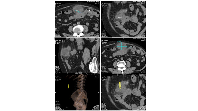
A 67 year-old male patient, presented with periumbilical and
left lumbar pain for one month that was not response to treatment.
Abdominal ultrasound detected one mixed echogenic mass
in the left lumbar mesentery with the diameter of 84 x 47mm. The conclusion was:
Suspected mesenteric infarction (Differential diagnosis: Intra-abdominal
Abscess) – Hepatic Steatosis – Abdominal Aortic Atherosclerosis.
Conclusion: Physicians should be on high alert when patients with abdominal pain not responding to the treatment. Abdominal ultrasound and MSCT help guiding the appropriate diagnosis for the case.
CASE 650: SOFT TISSUE TUMOR, Dr PHAN THANH HẢI, Dr JASMINE THANH XUÂN, Dr LÊ THÔNG LƯU, MEDIC MEDICAL CENTER, HCMC, VIETNAM
A 52 year-old patient with a mass at her right frontal region, no pain, no changed skin color.
Tuesday, 27 September 2022
CASE 649: CHOLANGIOCARCINOMA, Dr PHAN THANH HẢI, Dr VÕ THỊ THANH THẢO, Dr TRẦN CHÍ ĐỦ, Dr NGUYỄN THÀNH ĐĂNG, MEDIC MEDICAL CENTER, HCMC, VIET NAM.
A 68 year-old male patient with jaundice in ruling out of head of pancreas tumor from Tra vinh province went to Medic Center for an exact diagnosis before a surgical treatment.
Ultrasound ruled out pancreatic tumor and noted intrahepatic bile duct dilatation and 2 sides pleuresia. Gallbladder was not big with some sludge. Wall of bile duct thickening # 6-7mm, hyperechoic, CBD diameter 1.24mm existed 0.66--1.2 mm bile sludge at the beginning of CBD. Sign of bile duct tumor was unclear.
Surgery was done that detected bile duct tumor #1x2 cm. Removed bile duct tumor, GB, and connecting hepatic duct to jejunum by Roux-en-Y. Anapath was CholangioCarcinoma grade 2 invasive.
Tuesday, 6 September 2022
CASE 648 : SKIN ULTRASOUND, Dr PHAN THANH HẢI, Dr LÊ THANH LIÊM, MEDIC MEDICAL CENTER, HCMC , VIETNAM
A 90 year-old male patient with a painless node at right angle of his mouth. His past history was a left kidney cancer that had been cured for 5 years.
Skin utrasound by VINNO M86 system with linear probe 16MHz and 23MHz.
Focal cystic lesion # 9x4mm, represented mixed structure in subcutaneous layer, with fluid, non vascular, well limitted capsule. Elastography was green code that means a medium hardness. A benign mucous cyst was noted.
.jpg)


