Total Pageviews

Saturday, 17 June 2023

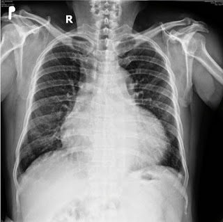
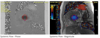
CASE 688: ASYMMETRICAL HYPERTROPHY of the HEART, Dr PHAN THANH HẢI, Dr NGUYỄN DINH, Dr ĐÀO XUÂN DUNG,MEDIC MEDICAL CENTER, HCMC VIETNAM.


A 59 year-old arterial hypertension male patient with a chest pain in taking alcohol from October 2019. He came Heart Institut HCMC with a note of infarctus of myocardium from Cantho province.  


But MSCT in Hoan my hospital Cantho province proves a slight stenosis 30% of coronary arteries. 

In COVID 19 pandemic, he goes around the private clinics with his HTA status and his heart but nothing change.

In May to July 2022 he goes to Medic Can tho and then Medic Hoa Hao Center in HCMC.

An coronary CTA of this second time shows slight stenosis under 30% and slight pericardial effusion but notes a hypertrophic cardiomyopathy of his heart.


At last Cardiac MRI confirms an asymmetrical hypertrophy of the heart.


He gets well with the management of  hypertrophic cardiomyopathy.


REFERENCE


Thursday, 15 June 2023

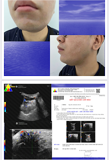
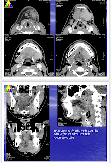
CASE 687: SUBMANDIBULAR TUMOR, Dr PHAN THANH HẢI, Dr TRẦN LÃM, MEDIC MEDICAL CENTER, HCMC, VIETNAM


 A 27 year-old male patient suffers from left submandibular gland for 3 years.

Ultrasound notes a left submadibular gland tumor.


MSCT confirms the left submandibular  that invaded the mouth floor and node.


But result of FNAC has only an amount epithelium cells and RBC.  And in the second time maybe a pleomorphic tumor.

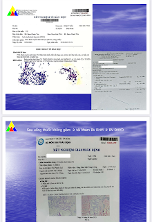
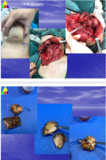
Goes through a surgery, the specimen is a malignant submandibular gland tumor, mucoepidermoid carcinoma low-grade.


Saturday, 10 June 2023

CASE 686: GASTRODUODENAL ARTERIAL ANEURYSM, Dr PHAN THANH HẢI, Dr PHAN THỊ HƯỜNG, Dr HỒ KHÁNH ĐỨC, MEDIC MEDICAL CENTER, HCMC, VIETNAM.


A 64 year-old HTA diabetic male patient is detected an arterial aneurysm # 35x34 mm by ultrasound maybe a gastroduodenal arterial aneurysm.


MSCT later confirms the 35x32 mm arterial aneurysm of gastroduodenal artery.


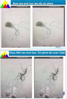
The patient goes through an endovascular aneurysm repair (EVAR) by biological glue.

The patient remains well post-op.

But ultrasound could note the vestige of aneurysm # 34x31 mm without any Doppler signal and flow after 8 months.

Wednesday, 7 June 2023

CASE 685: ANEURYSM of LEFT ILIAC VEIN and CORKSCREW of LEFT EXTERNAL ILIAC ARTERY, Dr PHAN THANH HẢI, Dr NGUYỄN ĐỨC DUY LINH, Dr NGUYỄN NGỌC XUÂN GIANG, BSc TRƯƠNG TẤN PHÁT, MEDIC MEDICAL CENTER and BINH AN HOSPITAL, KIEN GIANG PROVINCE, VIETNAM.

 A 58 year-old male patient in general check-up is revealed incidentaĺly by ultrasound an aneurysm of left iliac vein  # 48x66 mm without thrombosis. In addition, the left external iliac artery dilates #15-21mm appears in tortuosity like a corkscrew.


MSCT 64 confirmes the findings of the aneurysm of left iliac vein  # 48x66 mm without thrombosis. 


And in 3 D reconstruction the corkscrew of left external iliac artery appears clearly in mild dilatation without damage of its wall beside the aneurysm of the left iliac vein.

The corkscrew of external iliac artery is an anatomic variant incidentally revealed by CTA but a skilled sonologist could detects it with experience oneself. However, the key of this case is the aneurysm of iliac vein which leads to find out an uncomplicated aneurysm and the arterial tortuosity of the external iliac artery close by. But the cause of the left iliac vein aneurysm is unknown.

Aneurysm of iliac vein is a rare entity which appears in men and on the left side. And in contrast, female gender is a predisposing factor of the arterial tortuosity.


The patient is planned to the conservative management.

REFERENCES


:


Saturday, 27 May 2023

CASE 684: PRIMARY BREAST SARCOMA, Dr PHAN THANH HẢI, Dr LÊ THÔNG LƯU, MEDIC MEDICAL CENTER, HCMC, VIETNAM

A 61 year-old female patient suffers from 2 tumors of her right breast which are abandoned for 2 years by a deadly illness of her husband.

Ultrasound and elastography technique represent two BI-RADS 4B tumors of her right breast which one has perivascular signals.


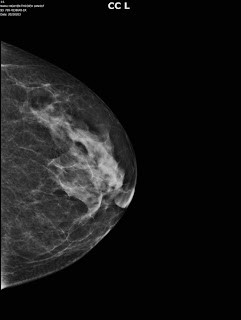
Mammography notes an asymetric sign in superiolateral region of the right breast.


MRI confirms the two BI-RADS 5 right malignant breast tumors: # 34x28 mm and # 21x 27 mm, with spiculated border, high signals on T2W2 and moderate signals on T1W1, captured contrast media type 3.


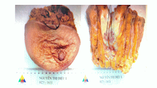
But the report of histoimmunology of the breast tumor is a breast sarcoma while axillary lymph nodes are not in malignancy.


Surgery is done in large field, no mastectomy nor lymph node curetage due to the sarcoma tumor characters.


As no clue of gene mutation, the patient goes through a planning of radiation therapy  for 3 months of 54 Gy dosages in 27 times.


DISCUSSIONS:

Breast sarcome is a rare mesenchymal breast tumor (<1% cancer breast tumor). MRI,  mammography and ultrasound could not differentiaze breast sarcoma from other breast cancer tumors.
Core biopsy and histoimmunologic exam are keys of diagnosis.
Surgery could save patient life that sarcoma invades in situ and rarely goes far via the blood stream. Chemotherapy and radiation may be managed in case of metastase and spreading exist. Liver, lung, bone marrow and recurrent breast tumor may happen in the first 2 years.  The 5-year survival rate reported in the literature ranges from 50% to 64% for the breast sarcoma.

The female patient remains well and in schedule of reexamination.

REFERENCES:

A rare case report of breast sarcoma - PMC-NCBI.

Primary breast sarcoma: case report-African Journal online.

Breast sarcoma: a case report and review of literature.


Saturday, 20 May 2023

CASE 683: A TB CASE: Dr PHAN THANH HẢI, Dr TRẦN THỊ TRÚC PHƯƠNG, MEDIC MEDIC CENTER, HCMC, VIỆT NAM

 A 29 year-old female  patient in a general chek-up for a preparation of a study abroad.

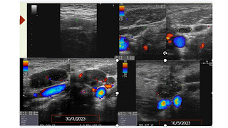
Ultrasound detects lymph nodes in her mesentery in the abdomen and on her neck # 5-25 mm in suspecting TB infiltrating type. Also a slight thickening of wall of colon is noted.


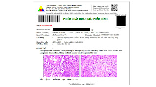
Report of the neck lymph node full biopsy is an TB  infected node.


A colonoendoscopy performs shows findings of ulcerative colitis from the cecum to the transverse colon due to TB infected.


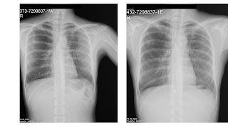
A TB regimen is planned for the patient, and during 2 months of TB management, her status is getting better, that is proven by the clear chest X-Ray and decreasing of lymph node in size and numeration.