Total Pageviews
Saturday, 23 July 2022
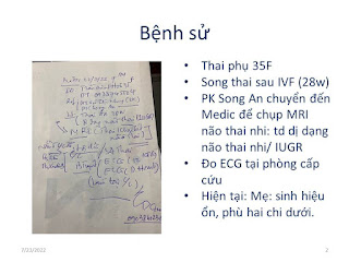
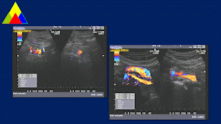
CASE 645: ULTRASOUND BUTTERFLY in EMERGENCY ROOM, MEDIC MEDICAL CENTER , HCMC, VIETNAM
Thursday, 21 July 2022
CASE 644: HEPATIC ABSCESS due to PERFORATED CHOLECYSTITIS with STONES, Dr DƯƠNG NGỌC THÀNH, Dr LÊ THANH LIÊM, Dr NGUYỄN HỮU QUỐC, Dr TRẦN THỊ HỒNG VÂN, Dr VÕ NG THÀNH NHÂN, Dr PHAN THANH HẢI, MEDIC MEDICAL CENTER, HCMC, VIETNAM
A 56 year-old female patient suffered from epigastric pain for one month with out fever. WBC 18,880/L , hCRP 129.5mg/L.
Ultrasound of abdomen revealed an abscess of liver that came from a cholecystitis due to stone. Gall bladder wall thickening was perforated that connected to the abscess. And there were stones in CBD and cystic duct.
MRI was done before a surgical treatment.
Endoscopic surgery at first but it did to change to open surgery to take away the hepatic abscess, that was in GB bed. Inflammed GB adhered to liver, mesentery and duodenum that has been dissected difficultly.
Surgeons performed partial cholecystectomy, and Kehr drainage after removing stones in CBD and in cystic duct.
DISCUSSION and CONCLUSION:Hepatic abscess due to perforated cholecystitis with biliary stone is still a rare entity. Ultrasound could detect successfully cholecystitis due to biliary stone [84-97 % sensitive, and 95-97% specific] that seems to be higher than CT or MRI does.
Saturday, 16 July 2022
CASE 643: MUCUS SECRETION versus ENDOLUMINAL TRACHEA TUMOR, LÊ HỮU LINH MD, MEDIC MEDICAL HCMC, VIETNAM
A 63 year-old male patient goes to the doctor as bloody sputum coughing for some days with feeling of suffocation. There was no pathological sign or symptom of other organs. Inflammed blood tests, and coagulation tests were in normal range.
His wife and his familial doctor also want having a bronchoscopy for the patient at MEDIC.
3 days later, patient no longer spitted bloody
sputum, and completely seems to be healthy. The pulmonologist decided to repeat chest CT and virtual bronchoscopy for the patient. Chest CT at MEDIC showed tracheal
lesion disappeared.
DISCUSSION:
Characteristics to detect a mucus
secretion in trachea:
• Small
size.
• At
posterior wall of trachea (supine position during CT scan procedure).
• Small
air shadow within the nodule suggesting mucus secretion.
• It
will be transformed or disappeared after coughing.
CONCLUSION:
Mucus secretion in trachea can be mistaken with endoluminal
trachea tumor.
Their characteristics on chest CT should be put under caution. And a repeated CT scan made after coughing may help detecting a mucus secretion. Thanks of that we could avoid unnecessary invasive procedure for patient.
A case with chest CT showed multiple nodular
lesions that were both upper lobes, indicated a pulmonary tuberculosis. And it existed a small
nodule at posterior wall of trachea. After coughing, repeated CT scan showed the nodule of trachea disappeared.
Friday, 15 July 2022
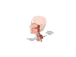
CASE 642: ZENKER' S DIVERTICULUM, Dr PHAN THANH HẢI, Dr NGUYỄN TUẤN CƯỜNG, Dr LÊ HỮU LINH, MEDIC MEDICAL CENTER, HCMC, VIETNAM
A 67 year-old male patient with multinodular goiter in reexamination.
Ultrasound Medic revealed a cystic lesion with air inside that was in posterior of right thyroid lobe. The lesion may be mimicking a calcified thyroid nodule in right lobe.
CT with CE confirmed the cyst captured contrast media which is in connection to esophagus on the right side. A Zenker's diverticulum was noted.
Thursday, 7 July 2022
CASE 641: SMALL BREAST TUMOR, Dr JASMINE THANH XUÂN, Dr PHAN THANH HẢI, Dr NẠI THỊ HƯƠNG THOANG, Dr TRẦN THỊ HỒNG VÂN, Dr HỒ CHÍ TRUNG, MEDIC MEDICAL CENTER, HCMC, VIETNAM
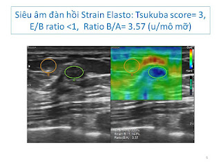
Small size breast tumor <10mm may be revealed early in yearly screenning.
Saturday, 25 June 2022
CASE 640: ISOLATED COMMON ILIAC ARTERY DISSECTION, Dr Lê Văn Tài, Dr Võ Nguyễn Thành Nhân, Dr Nguyễn Tuấn Vũ, Dr Hồ Khánh Đức, Dr Phan Thanh Hải, MEDIC MEDICAL CENTER, HCMC, VIETNAM
A 66 year-old male patient (170 cm height, 64 kg weight) with thoracic pain was suffering from arterial hypertension [164/84 mmHg, BP 64b/min] for 3 years, and diabetes mellitus for 2 years.
Ultrasound detected aliasing artifact from right common iliac artery [r CIA] which has been a dissection aneurysm # 34x23x20mm with 2 lumens. The peak systolic velocity (PSV) of the false lumen of right CIA was lower than the true one. It existed atheromatic plaques in the abdominal artery [AA] wall, but it [AA] was not aneurysm. A note of arterial aneurysm dissection of the right common iliac artery [r CIA] was made.
MSCT confirmed the 19x7 mm isolated right common iliac artery dissecting.
DISCUSSION and CONCLUSIONS
Isolated common iliac artery dissection is a very rare disease which is detected by chance. The possible causes included atherosclerosis, fibromuscular dysplasia, connective tissue disease, trauma and pregnancy.
It is certainly wherever to look for an arterial aneurysm in a hypertensive old patient in routine workflow not only for abdominal aorta (AAA) but also for other arteries and its branches or inside any abdominal organes.
Friday, 17 June 2022
CASE 637, 638, 639 : SECOND OPINION * X-RAYS-CT-ULTRASOUND, Dr PHAN THANH HẢI, Dr NGUYỄN VĂN CÔNG, Dr HỒ CHÍ TRUNG, Dr TRẦN NGÂN CHÂU, Dr LÊ THANH LIÊM, Dr LÝ VĂN PHÁI, Dr LÊ HỮU LINH, MEDIC MEDICAL CENTER HCMC, VIETNAM.
DISCUSSIONS
Case 1= Pneumomediastinum
Case 2= Fracture of left clavicule
Case 3 = Sealed - off right pleural effusion
Ultrasound could find out the abnormalities of 3 cases above but it need to confirm the diagnosing for them by other imaging modalities.


