Female patient 70yo with dysuria but without hematuria.
Ultrasound detected left kidney hydronephrosis grade 2 as a hyperechoic mass # 47x35mm inside renal pelvis that suggested a transitional cell cancer (TCC).


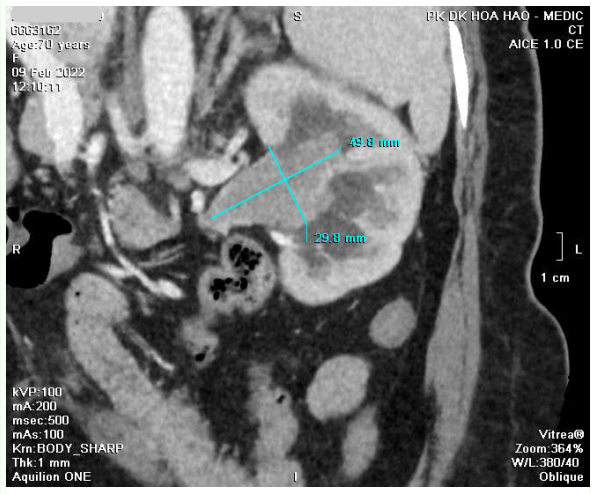
CT Scan: Soft tissue mass was in renal pelvis and ureter, d= 30 x 50 mm that highly captured contrast media while left kidney was in poor secretion of contrast. CT confirmed a left TCC.

It existed red and white blood cells and bacteria in urine analysis.
Endoscopic biopsy results was high malignancy uroendothelial carcinoma invaded the renal stroma.

Surgery removed left kidney and ureter. In longitudinal section of kidney, left pelvic kidney tumor sized # 5cm which was a necrotic vegetative mass while ureter was intact.

Pathological results : Transitional cell carcinoma poorly differentiated invaded parenchymal kidney. Non existed malignant cell in lymph nodes.