Tuesday, 29 August 2023

CASE 697: STOMACH GIST, Dr PHAN THANH HẢI, Dr PHAN THỊ HƯỜNG, MEDIC MEDICAL CENTER, HCMC, VIETNAM.

 A 47 year-old female patient with a gastric tumor at lesser curvature which was incidentally revealed by ultrasound.

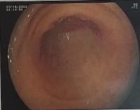
But gastroendoscopy result was only a gastritis.


Later MSCT confirmed a 50x58 mm gastric wall tumor maybe a stomach GIST. The tumor captured CE inhomogeneously and grew out of its way to compress the other organs around in abdominal cavity.


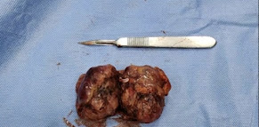
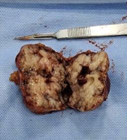
Robot surgery removed entirely the gastric tumor and recreated the cardia. The histopathological result is gastric GIST.


REFERENCE:


A 53 yearale patient with a gastric tumor which is spontaneously evealed 


Tuesday, 22 August 2023

CASE 696: PRIMARY CLEAR CELL HCC, Dr PHAN THANH HẢI, Dr NGÔ THỊ HUYỀN TRANG, MEDIC MEDICAL CENTER, HCMC, VIETNAM


A male patient 53 year-old with tumor in right lobe of liver and negative WAKO tests.

A 55 x 42 mm liver tumor is observed on ultrasound in segment 7. It is almost uniform, well-limited, weakly vascularized, and exhibits elastography ultrasonography SWE that is five times harder than hepatic parenchyma, measuring 29 kPa as opposed to 6.3 kPa. HCC risk testing is negative with WAKO.


MRI with Primovist confirms a 50 milimeter clear cell HCC (CC-HCC). T2 CE captured signals are higher than liver parenchyma and  lower than on T1. 


Biopsy results of tumor is an HCC well differentiazed.


REFERENCE:


Hepatocellular carcinoma (HCC) is a common cancer world-wide with a higher incidence in Asia. Clear cell variant of HCC (CC-HCC) has a frequency ranging from 0.4% to 37%. The presence of 90-100% clear cells is rare.


Saturday, 12 August 2023

CASE 695: Fasciola sp, UNUSUAL CAUSE of THICKENING of G I TRACT, Dr PHAN THANH HẢI, Dr TRẦN NGÂN CHÂU, Dr LÊ ĐÌNH VĨNH PHÚC, MEDIC MEDICAL CENTER, HCMC, VIETNAM.


Two cases of infected Fasciola sp whose larva migrants having in the same time some hepatic lesions and thickening of duodenum (2023) and right colon wall (2017) that are noted at Medic Center. 

CASE ONE:  A male patient 45 year-old, with history of thyroid cancer in 2013, enters a hospital as nausea, abdominal pain without fever after a ceremony buffet one day before.  Ultrasound detects hepatic lesions, and MRI  later reveals lesions in caudate lobe of liver and duodenum D3 wall thickening that is thought a case of infiltration of lymphoma on GI tract and liver.

But lab data notes raised highly the eosinophil proportion (48%) and positive Elisa tests for Fasciola sp and Gnathostoma.
Ultrasound of Medic Center confirmes liver lesions  and  duodenum D3 wall thickening  that maybe concludes due to infected parasites.


After 6 weeks managed by medical parasite drugs for Fasciola sp the male patient remains well; liver lesions reduce its sizes and duodenum wall gets normal on ultrasound and abdominal MSCT , and getting downed the eosinophil proportion.


CASE TWO: 

A female patient with Fascioliasis lesions in her liver and her right colon wall thickening in the same time which were detected by ultrasound and MSCT. 


Endoscopic biopsy of colon result was epithelial inflammation with eosinophil white blood cells.


She was managed successfully as Fasciola visceral larva migrants.
Larva migrants, especially for Fasciola sp, have a classic site in liver and biliary tree in acute phase and chronic phase. 
It exists two cases of larva migrants going outside the liver and biliary tree, to GI tract. If having lesions of it in liver or biliary tree one should not forget to find out larva migrants in another place.

REFERENCES:

https://www.nguyenthienhung.com/2008/10/c-im-siu-m-tn-thng-gan-do-sn-l-gan-ln.html?m=1

Ultrasound and Fascioliasis at MEDIC CENTER, Vietnam (slideshare.net) Oct, 21, 2013


Thursday, 10 August 2023

CASE 694: SPONTANEOUS SPLEEN LACERATION, Dr PHAN THANH HẢI, Dr NGUYỄN NGHIỆP VĂN, MEDIC MEDICAL CENTER, HCMC, VIETNAM.

A male patient 56 year-old  with one day of pain at left side of abdomen, obesity, and five-year chronic leukemia.  His history is noted splenohepatomegaly and cirrhosis and controlled type 2 diabetes. 

At Medic Center ultrasound detects free fluid in abdomen and big spleen with hematoma inside which makes thought about a spontaneous spleen laceration without any injured causes in spite of spleen infarction.

MSCT  with CE confirmes a spleen rupture with subcapsular hematoma and free fluid in the abdomen.


Surgery in emergency is done to remove the # 180x120x80 mm spleen and hemostasis procedure.
At the middle of anteromedial face of spleen it exists a #50 mm long  and 30 mm deep laceration line. Bloody fluid in abdominal cavity is an amount of 1.8 liter.
The patient goes through successfully the operation  but comes back with an 80x89 mm spleen bed abscess which is solved by medical management.


Chronic leukemia and obesity and DM may induce a spontaneous spleen laceration.

Thursday, 3 August 2023

CASE 693: ADVANCED MALIGNANT MESENCHYMAL CELLS of PROSTATIC TUMOR, Dr PHAN THANH HAI, Dr NGUYEN MINH THIEN, MEDIC MEDICAL CENTER. HCMC, VIETNAM.

 A 66 year-old male patient with weight loss and asthenia. Some 15-30 mm metastasized necrotic periaortic lymph nodes and prostate hypertrophy are noted on abdominal ultrasound.  Chest X-ray is normal and abdominal MSCT confirmes retroperitoneal lymph nodes and prostate tumor. PSA value is 1.38 ng/mL but with microscopic blood urine that transfers him to Medic Center for a prostate biopsy.

By via TRUS it exists a #55x43x62 mm big prostate, loss its capsule and distorsion of prostatic structure. The prostate tumor is noted having of many hard sites on shear wave ultrasound elastography.


Performed 12 specimen prostate biopsy and 16 items histoimmunopathologic report concludes a non- specialized mesenchymal cell of prostate tumor (PCa) on a chronic TB inflamed based structure.


This is a rare PCa malignant mesenchymal cell  of prostate tumor may happen on 1/1000 cases. The patient is waiting for an appropriate management due to his advanced status of the tumoral progress.