A 44 years-old male
patient, complaint of substernal chest pain for one week, increased with cough
and inspiration. He also had mild fever, dry cough, and dyspnea. He was first
seen on July 4th 2020, and was followed up at home with an initial
diagnosis of Suspected Pericarditis – Urinary Infection. He was then readmitted
7 days later at ER department.
ECG on July 4th,
2020 showed ST changes associated with pericardial effusion.
Blood test on July 4th, 2020 showed highly
elevated white blood counts, marked increase of hsCRP, and urinary infection.
The serum troponin was normal.
Echocardiography showed minimal pericardial
effusion.
Chest X-ray was normal.
He was given oral antibiotics (Levofloxacin) and anti-inflammation for 7 days.
On July 10th,
2020, he was admitted to ER due to severe chest pain, mild fever, and dyspnea.
Physical examination at ER showed tachycardia, normal BP, and no heart murmur.
Repeated ECG on July 10th, 2020 showed
flattened T-wave on DIII.
Second blood test
showed persistent elevated WBC, hsCRP and elevated D-dimers.
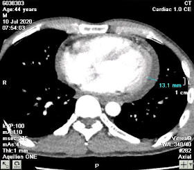
Chest CT-scan on July 10th, 2020 showed suspected mediastinum abscess surrounding the ascending aorta, with saccular aneurysm at the beginning of the aortic arch, and mild pericardial effusion. The differential diagnosis was thoracic aortic aneurysm with surrounding hematoma.
The patient was then
transferred to Binh Dan Hospital. He was operated on the very next day, and
surgery report showed inflammation and necrosis of the aortic aneurysm’s wall.
The necrotic tissues were removed, and the aortic arch was partially replaced
with a Vascutek 16 graft.
During his hospital staying, pericardial fluid culture
came back positive for Staphylococcus aureus. He was treated with a combination
of Vancomycin and Imipenem.
He’s currently stable with minimal pain at the
surgical site. His white blood count went down to almost the normal range.
CONCLUSION=
Echocardiography and EKG detected pericardial effusion, CT revealed infected aneurysm and mediastinal abscess and patient remained well post-op ; that is a great success for saving patient life came from an interesting combination of clinical and imaging of diagnosing and surgery.


No comments:
Post a Comment