X- Rays : Normal chest and vertebral column.
Blood tests : WBC ,
hs CRP : normal.
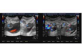
Ultrasound of pelvis:
Mass with mixed pattern of structure presses on urinary bladder that connects retroperitoneum and covers right poas muscle.
Mass goes forward under right pelvic wall and presses on peritoneum.
And enters muscle layers of right pelvic wall.
A diagnosis of pelvic abscess is made by sonologist.
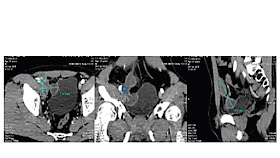
MSCT : Lesion in right pelvic wall#5x8cm, cystic , multicrescent, thick capsule with septation which takes contrast and presses urinary bladder and goes down to right inguinal canal. Radiologist thinks about a pelvic wall abscess.
MRI : Right pelvic abscess in retroperitoneum goes forward that presses on urinary bladder then goes upward to right pelvic wall muscles.
FNAC withdraws some milky fluid, like abscess fluid.
Core biopsy results TB pelvic abscess.
A 6 month TB planning is done for this patient.


No comments:
Post a Comment