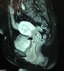
 Woman 48yo, PARA 2002 , detected herself one prolapsed mass from her vagina 1 year ago, no pain, no fever but with SUI [ stress urine incontinence] syndrome.
Ultrasound of pelvis by the transcutaneous: (US1) uterus normal size,
by via TVS ( US2, US 3) detected one mass at lateral left uterus, hypoechoic, size 7 cm look-liked second uterus.
CT scan of pelvis: CT 1 : this mass at left site uterus, hypodense like fatty tissue.
CT2, CT3 the mass is anterior the urinary bladder.
MRI 1, MRI 2 in sagittal section, this tumor is like a second uterus.
OPERATION REMOVE THIS TUMOR..BY LAPAROTOMY..
( OP. IMAGES:THIS TUMOR IS NOT FROM UTERUS)
MICROSCOPY IS FIBRO-LIPOMA


No comments:
Post a Comment