Friday, 23 October 2015

CASE 343: TB AXILLARY NODES, Dr PHAN THANH HẢI, Dr JASMINE THANH XUÂN, MEDIC MEDICAL CENTER, HCMC, VIETNAM.

FOR PICTURES PLS CONNECT TO 3 G / DOWNLOAD THE LINK

case-343-tb-axillary-nodes-

Woman 49 yo, on mammography screening detected  many  left axillary nodes and calcification, no  detected tumor in mammary gland (see mammogram).


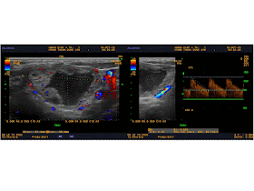
Ultrasound  of left   axillary  found  many  lymph nodes,   sizes of 1-2 cm,  round
and  calcification  inside node ( US picture 1). 


CDI cannot  detect  hilus  of nodes, no vascular signal  in  nodes, ( US 2, US 3). Elastoscan  US of this  node was hard,    17.3 kPa ( US 4)


MRI  with  FAST SCAN   DWI..made sure  no tumor intra  left  breast and  
axillary nodes.


Biopsy   removed one  big node with  structure  inside  look liked  caseum.


Microscopy result was   tuberculosis  with  typical  big cell  LANGHANS.


CONCLUSION: Tuberculosis of axillary lymph nodes..

Tuesday, 20 October 2015

CASE 342 : FRONTAL LUMP, Dr PHAN THANH HẢI, Dr LÊ THỐNG NHẤT, MEDIC MEDICAL CENTER, HCMC, VIETNAM

FOR PICTURES PLS CONNECT TO 3 G / DOWNLOAD THE LINK


MAN 65 YO  ONE YEAR AGO  DETECTED  AT  RIGHT FRONTAL A SMALL MASS UNDER SCALP  SLOWLY GROWING WITHOUT PAIN.


ULTRASOUND FINDINGS=

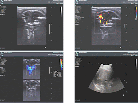
US 1:TUMOR  DESTRUCTING   FRONTAL BONE, SIZE OF 2CM.

US 2:  HYPERVASCULAR TUMOR.

US 3:ELASTOSCAN OF TUMOR OF 36.3kPA.

US 4.  ULTRASOUND FINDINGS OF  LIVER TUMOR OF 5 CM.


US 5: ELASTOSCAN  OF LIVER TUMORS = HARD,  55.9 kPA


MSCT  BRAIN=  3  CT PICTURES SUSPECTED  METASTASIS TO FRONTAL BONE.


BLOOD TESTS=   HCV POSITIVE  AND WAKO TEST  WASE  STRONG POSITIVE WITH DCP. IT MEANS  HCC.


FNAC  OF THE  FRONTAL TUMOR WAS  HCC METASTASIS.


CONCLUSION:  HCC IN LIVER  METASTASIS TO  FRONTAL BONE.

REFERENCE


Sunday, 18 October 2015

CASE 341: THYROID TOXIC ADENOMA, Dr LÊ TỰ PHÚC, MEDIC MEDICAL CENTER, HCMC, VIETNAM


A 41 yo male patient with chronic fatigue syndrome and  nervousness, irritability; sometimes  he feels muscle weakness and hand tremor  for three months and getting severe in ten days. Wants to check up his liver and nervous system. 

Abdominal ultrasound revealed nothing abnormal. Because of his symptoms, sonologist also perfomed a thyroid ultrasound.

Thyroid ultrasound showed that  right lobe and  upper portion of  left lobe were normal in size with smooth margin and homogeneous echotexture, normal blood flow in Doppler ultrasound.

But  lower portion of the left lobe had a 5 cm, mixed cystic-solid nodule with hypervascular, isoechoic in peripheric part and nonvascular cystic degeneration in center part of tumor.


On Doppler US,  inferior thyroid artery showed  peak systolic velocity in  left lobe is 122.7 cm/s, five times more than one of right lobe 24.3 cm/s. So,  sonologist suspected  nodule in  lower left lobe maybe a toxic thyroid adenoma, which is cause of  hyperthyroidism.


Blood tests were done and confirmed the diagnosis with low level of TSH and high level of Free T3, Free T4.


Measuring the peak systolic velocity of inferior thyroid artery in both side to diagnose toxic thyroid adenomaDo you think we can diagnose toxic thyroid adenoma by ultrasound?

Thursday, 15 October 2015

CASE 340 : UMBILICAL TUMOR, Dr PHAN THANH HẢI, Dr LÊ THÔNG LƯU, Dr NGUYỄN THị KIM UYÊN, MEDIC MEDICAL CENTER, HCMC, VIETNAM

FOR PICTURES PLS CONNECT TO 3G / DOWNLOAD THE LINK

Woman 45 yo, PARA 2002, normal spontaneous vaginal deliveries,no cesarean operation  nor history of hormonal contraception. 

2 years ago  she  detected  her umbilicus swelling some days before her menses and continuous pain  during  the entire of her period in some times bleeding.
In clinical  examination  the umbilicus  deformed  by one mass which were bluish-black, hard and not  hot (see 2 photos).


Ultrasound findings of this mass=
US1: this mass was well bordered, localized in cavity of  navel. Structure of mass was solid, size of  2.68 cm.


US 2:  in CDI, vascular supplying from peripheral part of mass.


US 3:  in elastography:  hard mass in comparison to muscle.


US 4 : uterus and pelvis were not intact.

Preoperative diagnosis is primary endometriosis. Removed this tumor. See macroscopic specimen.


Microscopic report: ENDOMETRIOSIS.


CONCLUSION: This  is  a case of   PRIMARY  ENDOMETRIOSIS in umbilicus.

REFERENCE:


Saturday, 10 October 2015

CASE 339: INTRAMUSCULAR TUMOR, Dr PHAN THANH HẢI, MEDIC MEDICAL CENTER, HCMC, VIETNAM.

FOR  PICTURES PLS CONNECT TO 3 G / DOWNLOAD THE LINK


Man 51 yo  detected  himself  swollen at  left  scapula region, no pain for 2 years (photo). 


The tumor  was  underskin and ovoid shape.
Ultrasound  of this mass was  localized   in  trapezius muscle, well bordered,  size of 10cm. CDI  no abnormal blood  flow. Elasto scan was  slow  kPa (7.7kPa) in  comparison to muscle 22.9 kPa.


MRI  scan= MRI 1,tumor  well bordered with  density as  fatty tissue, MRI 2, MRI 3).


Operation  was done  for removing of  the tumor.
Microscopic report of this tumor is LIPOMA.


Monday, 5 October 2015

CASE 338: THYROID CANCER, Dr PHAN THANH HẢI, MEDIC MEDICAL CENTER, HCMC, VIETNAM.

FOR PICTURES PLS CONNECT TO 3 G / DOWNLOAD THE LINK

case-338-thyroid-cancer


Man 30yo, in general check-up,  ultrasound detected  thyroid tumor  at right and left  lobes.
US1 scan at   right lobe, small nodule 1cm diameter,  hyperechoic due to  calcification.


US 2 scan at  left  lobe,    round border tumor, 4 cm with many white calcification spots.


US 3 & US 4:  CDI of  left  thyroid tumor, hypervascular.


US 5  elasto scan of right  tumor was very hard.


US 6 elastoscan with Q box score,  tumor in comparison to  normal thyroid tissue.


No detection of  regional lymph nodes.
Report  by  sonologist   was suspected  thyroid carcinoma, THYRADS IV, and FNAC of  the left tumor was  PAPILLARY  CARCINOMA.


DISCUSSION: B MODE  SCAN  THYROID TUMOR   WITH MANY  WHITE SPOTS  WITHOUT  SHADOWING, IT IS   MICROCALCIFICATION NAMED   PSAMMOMA BODY..WHICH  IS  TYPICAL  OF  PAPILLARY THYROID CARCINOMA.

ELASTOSCAN THIS TUMOR   WITH  QUANTITATIVE Q-BOX  IS 99.5 kPa  IN COMPARISON WITH   NORMAL THYROID  GLAND IS  12.2 kPa.
ELASTOSCAN  IS NEW  TECHNOLOGY  FOR  DETECTION  THYROID  CANCER.


REFERENCE