Monday, 11 February 2013

CASE 168: NOT HAVING GALLBLADDER, Dr PHAN THANH HẢI, MEDIC MEDICAL CENTER, HCMC, VIETNAM

Man 48 yo in hospital admission by fever and jaundice progressing. Physical examination: pain at right subcostal area, past history was known diabetes and gallbladder stone. Plain abdominal XRay film standing showed ilius status.


Ultrasound of abdomen revealed small fluid collection at the border of liver, and CANNOT FIND OUT THE GALL BLADDER BUT DETECTED ONE  HYPERECHOIC MASS ADHERED TO LOWER BORDER OF LIVER. THE CBD WAS NOT IN DILATATION.


Blood tests with elevated WBC of 16K (90% neutrophil).

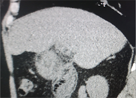
MDCT non CE found that the gallbladder without stone nor fluid into gallbladder.


What is your explanation of the ultrasound images for this gallbladder?


Based on clinical  status:  fever, jaundice, pain at right subcostal area, and imaging modalities (abdomen plain film, ultrasound  and MDCT) with  blood tests, the  diagnosis was acute cholecystitis lead to gallbladder empyema. The IV antibiotic resulted clinically good response in medical treatment.

Reference: 


No comments:

Post a Comment